Background
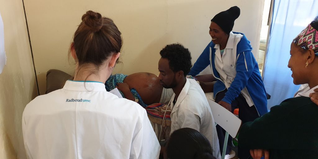
Ultrasound imaging is widely used for prenatal screening. Unfortunately, prenatal ultrasound imaging is barely performed in resource-limited settings. This is mainly caused by a severe shortage of well-trained personnel that is able to acquire and interpret ultrasound images. In this project, we are therefore combining Artificial Intelligence with a predefined ultrasound acquisition protocol. This acquisition protocol can be taught to a midwife within two hours. Combining this protocol with deep learning algorithms has the potential to obviate the need of a trained sonographer in resource-limited settings.
With the acquisition protocol, data was already acquired from 390 pregnant women in Ethiopia. This data will be used to develop two new deep learning algorithms: one algorithm that automatically detects twin pregnancies and the other algorithm that automatically determines the location of the placenta. Timely detection of twin pregnancies is crucial for delivery management. When the location of the placenta is known, it is possible to detect women that are at risk of placenta previa (which is a placenta localized close to the birth channel). Women with placenta previa have a two-fold increased risk of postpartum hemorrhage and even a ten-fold increased risk of antepartum hemorrhage.
Research question and tasks
Development of deep learning algorithms that automatically determines the location of the placenta and automatically detects twin pregnancies using a predefined ultrasound acquisition protocol.
Tasks
- Learn about ultrasound imaging and the acquisition protocol that is used
- Development of deep learning algorithms
- Evaluation of the developed algorithms
- Optimize the algorithms on computational efficiency
Innovation
When the algorithms have sufficient sensitivity and accuracy, and are able to run on a smartphone, they will be integrated in the current prototype which we are planning to evaluate in resource-limited settings.
Requirements
- Students in the final year of a study in computer science, artificial intelligence, physics, or a related area.
Information
- Project duration: 6 months
- Location: Radboud University Medical Center
- For more information, please contact Thomas van den Heuvel


