Background
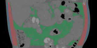
Body composition is an important biomarker in the treatment of cancer. In particular, low muscle mass has been associated with higher chemotherapy toxicity, shorter time to tumor progression, poorer surgical outcomes, impaired functional status, and shorter survival. However, because CT-based body composition assessment requires outlining the different tissues in the image, which is time-consuming, its practical value is currently limited. The different tissue types are often outlined manually and only in a single slice of a CT scan at the level of the third vertebra (L3). This is based on the observed linear relation between cross-sectional areas and total body volume of muscle and fat. However, 2D estimates are still less accurate than 3D measurements. For use in both routine care and in research studies, automatic segmentation of the different tissue types is desirable, especially if that would enable quantification of various tissue types in the whole 3D scan rather than a single slice.
Research question and tasks
Develop, train and test a deep learning algorithm that automatically outlines different muscles and fat in 3D CT scans. An important aspect of this project is also the implementation as an easy to use web-app so that the software can be used by clinical researchers.
Requirements
- Students in the final year of a Master study in computer science, artificial intelligence, biomedical engineering, or a related area.
- Good programming skills are important
Information
- Project duration: 6 months
- Location: Radboud University Medical Center
- For more information, please contact Nikolas Lessmann


